The 2026 Nobel Prize in Medicine: Why Controlling Neurons with Light Matters
How optogenetics works, what a causal experiment can tell us, what researchers have achieved, and why a research tool can deserve a Nobel Prize.

mou7s
Scientists can often see neurons becoming active without knowing exactly what those cells are doing. When an animal lifts a leg, a group of neurons becomes active. Did they issue the movement command, or did they receive feedback after the leg moved?
Optogenetics lets researchers change the activity of selected cells and observe the result. It moves an experiment beyond watching the brain work toward testing how it works.
The 2026 Nobel Prize in Physiology or Medicine recognizes Karl Deisseroth, Peter Hegemann and Georg Nagel for discoveries concerning light-gated ion channels and optogenetics. Award and contribution information
To understand the significance, follow one question: if we suspect that a group of neurons controls movement, how can we test it?
Are the cells sending a command or receiving a message?
Imagine observing a mouse. Every time it lifts a leg, a particular group of neurons becomes active.
The cells might be telling the body to move. They might be receiving news that movement has occurred. Or they might become active alongside the cells that actually control the movement. Watching these events happen together does not distinguish the explanations.
A more direct approach is to activate the cells ourselves and check whether movement changes. We can also suppress their activity temporarily and examine whether normal movement is affected.
That requires selecting the cells we want to study and controlling when and for how long we stimulate them. Optogenetics provides a way to design such experiments.
The mouse experiment below is a hypothetical teaching example, not a description of a particular study.
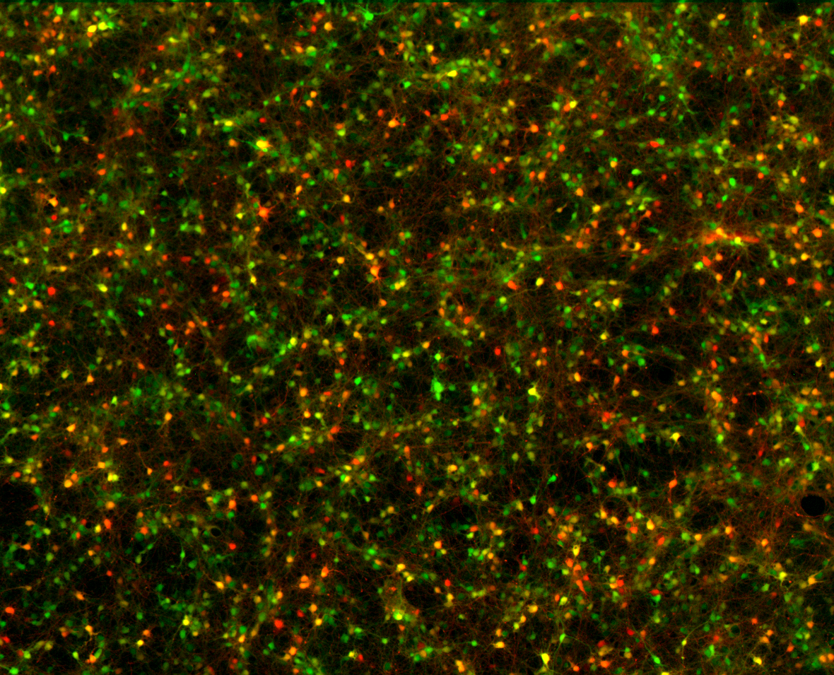
Cultured neurons under a fluorescence microscope. Red and green fluorescent proteins label expression patterns; this is not an image of neurons being activated by light. Image: ManuelSchottdorf / Wikimedia Commons, CC BY-SA 4.0, unmodified.

Step one: give selected cells a “light switch”
Ordinary neurons will not respond to a beam of light in the way an experiment requires. Researchers first give them a suitable light-sensitive protein.
They deliver a gene encoding the protein, rather like providing a manufacturing instruction. The cells use that instruction to make the protein and place it in their membranes.
Installing a switch therefore means getting a cell to produce a light-responsive molecule. This explains the name: genetic expression gives selected cells light sensitivity, and light changes their activity.
How does the switch affect a cell?
Neurons communicate through electrical signals. Voltage across the cell membrane is influenced by the movement of ions: electrically charged particles.
Think of the membrane as a wall and an ion channel as a gate. Some light-sensitive proteins act as gates that open under suitable illumination. Ions pass through and change the cell’s voltage.
A classic example is channelrhodopsin-2, or ChR2. A 2003 study established it as a directly light-gated cation channel. This discovery grew out of research into how microorganisms such as green algae respond to light. Original paper
When ChR2 operates in a neuron’s membrane, illumination can make electrical firing more likely. Other light-sensitive tools can suppress activity. The effect depends on the tool and the cellular conditions.
Step two: bring light to the cells
Once cells have the switch, light still needs to reach it.
Some animal experiments targeting deeper brain regions use a fine optical fiber positioned near the area of interest. It provides a route for light to reach the target.
There are two selections involved: which cells produce the protein, and where illumination reaches. Together, they help determine which cells are affected.
Researchers also control when the light turns on and off, coordinating stimulation with an experimental task. A landmark 2005 paper demonstrated millisecond-timescale optical control using ChR2 in neurons. Original paper
Boyden, Zhang, Bamberg, Nagel and Deisseroth collaborated on that paper. The laureates made important contributions, and the development of the method also involved other researchers.
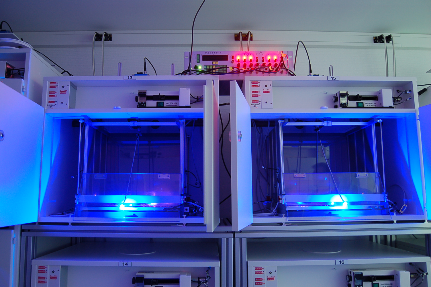
Equipment for optogenetic stimulation and behavioral studies in rats, showing cages and connections. This is an example of real equipment, not the hypothetical mouse experiment in the text. Image: Bd008 / Wikimedia Commons, CC BY-SA 4.0, unmodified.

Step three: change activity and observe movement
Return to the mouse example. Researchers compare movement without illumination against movement when light activates the selected neurons.
A change after activation provides evidence that the cells can influence movement. Temporarily reducing their activity with a suitable inhibitory tool can further test whether normal movement depends on them.
Two checks are essential.
The cells must respond as intended. A light turning on does not establish that neural activity changed. Measurements such as electrical recordings help verify the response.
The effect must be connected to the tool. For example, applying the same illumination to cells without the light-sensitive protein helps check whether light or the apparatus produces a similar effect on its own.
These checks connect a deliberate change with the outcome that follows.
Why is this stronger than watching?
The difference is straightforward:
| Method | What it reveals |
|---|---|
| Observation | These cells are active when the mouse moves |
| Intervention | Changing these cells’ activity changes movement |
The second result offers more direct causal evidence because researchers deliberately changed a condition. It still needs verification and controls.
Influencing movement also does not mean exclusively controlling movement. The brain is interconnected, and the same cells may contribute to several tasks. A result tells us what those cells do under the conditions of that experiment.
The switch analogy explains the tool. It does not imply that the brain is a row of switches, each responsible for exactly one function.
What has the method achieved?
Its most immediate achievement is an experimental capability. Researchers can test relationships between selected cells and behavior more directly, applying the approach to questions about movement, memory and emotion.
A tool can therefore matter across many laboratories and many questions. Its impact grows beyond its first demonstration.
A human application offers a more tangible example: partial visual recovery in a blind patient.
Giving surviving retinal cells a way to sense light
In 2021, Sahel, Roska and colleagues reported an experimental treatment in a patient with retinitis pigmentosa, a disease that damages photoreceptors and can lead to blindness. Study abstract
Some surviving retinal ganglion cells were made to produce the light-sensitive protein ChrimsonR. Engineered goggles converted visual information into light signals projected onto the retina, stimulating the modified cells.
The idea was to give remaining cells part of the light-sensing role after the original photoreceptors were damaged.
With the treated eye and goggles, the patient could perceive, locate, count and touch some objects. Without the goggles, the same object-detection ability was not demonstrated.
This was partial recovery, not normal vision. A single case cannot establish the same benefit for every patient. It nevertheless demonstrated a possible route toward restoring some function by changing how cells respond to light.
Another team conducted this clinical research. It illustrates a later application of optogenetics and differs from the brain-fiber experiment described above in both equipment and target cells.
Why can this deserve a Nobel Prize?
The significance becomes clearer after following the experiment. Optogenetics enables researchers to select cells, change their activity at a chosen time, and test their role.
I find the microscope a useful comparison. Its value extends beyond what the first user saw: it lets later researchers examine things previously inaccessible to them. An experimental tool such as optogenetics similarly supports many different investigations.
This is an interpretation of the scientific importance. The contribution information is linked at the beginning of the article.
The starting question is also worth considering: how do microorganisms respond to light? It seems distant from the human brain, yet it helped produce a tool for studying nervous systems. The consequences of basic research can emerge gradually through years of work by many people.
Understanding the award
Medical progress naturally raises the question, “Which disease has it treated?” That matters. Optogenetics also invites another question: what can we now investigate more accurately?
Understanding how cells and pathways work improves our ability to ask where disease disrupts a system and how an intervention might help. Research tools can therefore be part of medical progress.
Moving from an experimental tool to everyday treatment still requires work on delivery, equipment, long-term outcomes and safety. Animal experiments, individual human reports and established treatments represent different stages of evidence.
The capability worth remembering is this: light can change selected cells’ activity, allowing explanations about the brain to face more direct experimental tests.
Sources checked on 6 October 2026. The movement experiment is hypothetical; the vision case is a published study. The cover is a conceptual illustration.
Sources and further reading
The 2026 Nobel Prize in Literature: How Anne Carson Makes Ancient Stories Hurt Again
A guide to Anne Carson through a modern monster’s coming of age, the bittersweet experience of desire, and mourning expressed through an accordion-fold book.
The 2026 Nobel Prize in Physics: Why Bury a Telescope in Antarctic Ice?
Why neutrinos are difficult to detect, how light in ice reveals their interactions, and what IceCube has achieved as a new way to observe the universe.